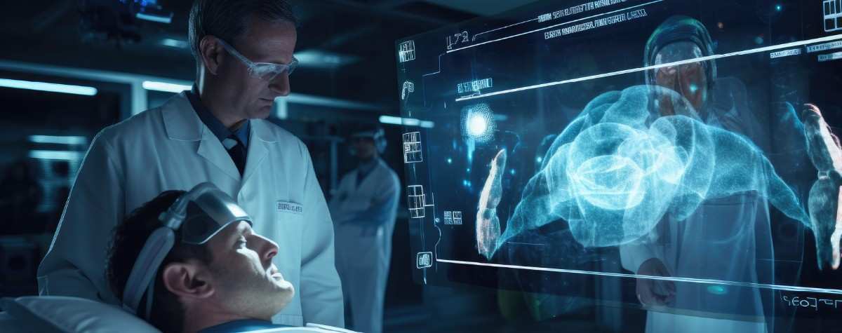


Improve diagnostics with AI-driven medical imaging solutions. Enhance accuracy, streamline workflows, and enable early disease detection.
Medical imaging solutions are essential for making accurate and quick diagnoses. This is central to quality healthcare. Imaging technology has progressed from X-rays to advanced AI-based imaging. These have the ability to diagnose diseases at an earlier stage and with better accuracy. These technologies improve diagnosis now and shape future patient care.
Modern technologies, like MRI and CT scans, help doctors work faster. Real-time 3D imaging and predictive analytics give them better information. This helps in making informed decisions. In this blog, we will look at how these technologies are changing diagnostics. They enhance treatment results and what we can do in healthcare.
Medical imaging solutions have changed a lot since scientists discovered X-rays in 1895. We’ve moved from grainy black-and-white images of bones to high-resolution, AI-powered scans. These advancements help find diseases early. They have boosted diagnostic accuracy. This change has transformed how healthcare providers treat and care for patients.
The journey started with X-rays. This was a revolutionary discovery. It helped doctors view the inside of the human body without surgery. Progressively, breakthroughs such as ultrasound, CT scans, and MRI produced even more detailed pictures. They depicted soft tissues, organs, and blood flow in real time. PET scans built on this further. They enabled doctors to see metabolic activity. This shift influences the way doctors detect and treat cancer.
The real shift came with digital imaging and artificial intelligence. AI-assisted imaging helps radiologists identify abnormalities with greater speed and precision. This reduces errors and boosts early detection rates. Machine learning algorithms scan thousands of images in seconds. They find patterns that human eyes might miss. This accuracy leads to better therapies and improved patient outcomes.
X-ray images show fractures, infections, and lung disorders. CT scans create cross-sectional images. These assist in identifying tumors, internal hemorrhages, and vascular diseases. AI-driven analysis enhances interpretation, reducing the error rate and improving efficiency.
MRI uses radio waves and magnetics to develop clean images of soft tissues. It assists physicians in diagnosing neurological and musculoskeletal problems. Ultrasound provides real-time images for fetal monitoring, heart screening, and organ testing. AI software enhances image quality and detects abnormalities automatically.
Positron Emission Tomography (PET) scans use radiotracers to track metabolic activity. This helps in early cancer detection and neurological exams. Single-Photon Emission Computed Tomography (SPECT) imaging checks blood flow and organ function. It also aids in diagnosing heart and bone diseases. AI-based image reconstruction improves scan quality and speed.
AI in medical imaging uses data from sources like NIH ChestX-ray14, LIDC-IDRI, and BraTS. They look for patterns in the data. AI preprocessing like artifact and noise elimination enhances the clearness of an image. Anomaly detection and segmentation are enhanced by Convolutional Neural Networks (CNNs) and transformers. These tools speed up diagnosis and reduce manual review time.
Advanced medical imaging solutions depend on structured, high-quality data for precise analysis. Techniques such as normalization and contrast adjustments optimize imaging input for AI models. Real-time interpretation supports clinicians in making faster, more informed decisions. Scalable cloud-based AI deployment ensures efficient processing and regulatory compliance.
AI extracts imaging features through deep learning, reducing human subjectivity in diagnosis. CNNs examine LIDC-IDRI and BraTS scans. This helps boost the detection of lung nodules and brain tumors. Federated learning allows AI to train institutions without compromising data privacy. Smart workflow automation prioritizes urgent cases and lessens radiologists’ workload.
AI spots disease trends by analyzing past imaging data. Predictive algorithms from LIDC-IDRI and NIH ChestX-ray14 can detect early signs of diseases. They identify issues like lung cancer and pneumonia. AI assessments target high-risk cases before symptoms show. Continuous learning improves diagnostic accuracy and responsiveness for various patient groups.
AI links imaging results to lab data, patient history, and EHRs. This improves diagnostics. FHIR and DICOM-compliant APIs enable smooth system integration and real-time HL7 messaging. Blockchain audit trails enhance data security and ensure compliance. Cloud-based AI allows for real-time diagnostics at reduced infrastructure costs.
